Exploring Cancer Immunotherapy: Understanding the Mechanisms of Therapeutic Antibodies
View pictures in App save up to 80% data.
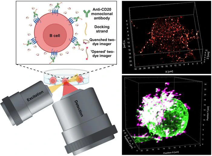
In the field of medical research, an innovative advancement has emerged from the University of Würzburg's laboratories in Bavaria, Germany. The study led by Professor Markus Sauer and his team represents a significant progression in the comprehension and creation of therapeutic antibodies. This research sheds light on the complex interactions between antibodies and their target molecules, especially in relation to B cells linked to blood cancers, including chronic lymphocytic leukaemia. The research team has pioneered a cutting-edge super-resolution microscopy method called LLS-TDI-DNA-PAINT, which enables real-time visualization of therapeutic antibodies attaching to target proteins on cancer cells in three-dimensional space, achieving an unprecedented level of detail and precision.
For many years, immunotherapeutic antibodies have played a crucial role in the treatment of different types of tumors. A key focus of these therapies is the CD20 protein found on the surface of B cells. The binding of these antibodies to CD20 triggers a series of immune responses that ultimately result in the elimination of cancerous cells. Nonetheless, even with the proven effectiveness of these antibodies in clinical settings, scientists have struggled to fully understand how they bind and the cellular responses that follow. Recent groundbreaking research from Würzburg has made significant strides in addressing this knowledge gap.
Using the LLS-TDI-DNA-PAINT method, scientists successfully observed the real-time dynamics of therapeutic antibodies. They examined the interaction between these antibodies and CD20 molecules, focusing on how this binding affects the structural behavior of B cells during the process. A significant finding from the study was the observation of a phenotypic change in B cells, which the researchers referred to as adopting a “hedgehog shape.” This alteration occurs due to the crosslinking of CD20 proteins by the antibodies, which activates complement systems and enhances targeted immune responses.
The researchers performed their observations utilizing the Raji B cell line, which is derived from human Burkitt's lymphoma and is well-regarded for its significance in cancer research. This specific cell line acts as a model for evaluating B cell responses to therapeutic antibodies. The research team executed experiments with four different therapeutic antibodies: RTX, OFA, OBZ, and 2H7, all of which are recognized for their targeting of CD20. The findings from their studies indicated that, irrespective of the antibody type used—whether categorized as type I or II—the response mechanism was remarkably similar, challenging earlier beliefs held by the scientific community.
Additionally, research has demonstrated that therapeutic antibodies primarily bind to specific areas of the B cell membrane, particularly at microvilli.